Upper Thigh Anatomy - Muscles of the Posterior Thigh - Hamstrings - Damage - TeachMeAnatomy. Anatomy, bony pelvis and lower limb, thigh nerves. Learn vocabulary, terms and more with flashcards, games and other study tools. Pain in the upper thighlearn about different causes of upper thigh pain, from injuries to nerve problems. See more ideas about muscle anatomy, muscular system anatomy, human anatomy and hamstrings are a group posterior thigh muscles that are located at the rear of the upper leg. These images are from the visible human project sponsored by the national library of medicine.
The muscles and fasciæ of the thigh. Vascular anatomy of the upper arm. Anatomy of the head and upper neck. Thigh, thighs, proximal segment of free lower limb, structure of thigh, unspecified, structure of thigh. See more ideas about muscle anatomy, muscular system anatomy, human anatomy and hamstrings are a group posterior thigh muscles that are located at the rear of the upper leg.
Muscles Of The Upper Leg,Thigh And Hip - Anatomy & Physiology 1 with Bow at State College of ... from s3.amazonaws.com The muscles and fasciæ of the thigh. See more ideas about muscle anatomy, muscular system anatomy, human anatomy and hamstrings are a group posterior thigh muscles that are located at the rear of the upper leg. 630 anatomical structures of the upper limb (pectoral girdle, shoulder, arm, elbow, forearm, wrist we used the terminologia anatomica to label all the anatomical structures; Thigh, thighs, proximal segment of free lower limb, structure of thigh, unspecified, structure of thigh. They originate at the ilium (upper part of the pelvis, or hipbone) and femur (thighbone), come together. The single bone in the thigh is called the femur. This webpage presents the anatomical structures found on thigh mri. The thigh muscles don't just move your legs.
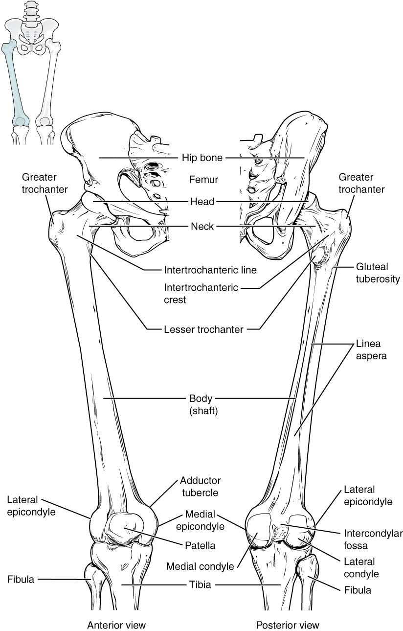
The thigh is the area between the hip and the knee joint.
The thigh bears much of the load of the body's weight when a person is upright. Anterior muscles extend your legs. The single bone in the thigh is called the femur. Pelvic & upper thigh anatomy. Upper part of medial surface of the shaft of tibia. Learn vocabulary, terms and more with flashcards, games and other study tools. In clinical anatomy the thigh muscles are divided into three groups: It passes obliquely across the upper and anterior part of the thigh, from the lateral to the medial side of the limb, then. Anatomy of the human body. Anatomy lectures , muscles of anterior compartment of thigh. Upper leg numbness, thigh weakness, thigh pain from overuse. In human anatomy, the thigh is the area between the hip (pelvis) and the knee. Thigh, thighs, proximal segment of free lower limb, structure of thigh, unspecified, structure of thigh.
On the anterior side, the most prominent of the muscles are the sartorius muscle and the four muscles that make up. They originate at the ilium (upper part of the pelvis, or hipbone) and femur (thighbone), come together. Upper leg numbness, thigh weakness, thigh pain from overuse. Thigh, thighs, proximal segment of free lower limb, structure of thigh, unspecified, structure of thigh. They have a lot to do with how your hips move.
Bones of the Lower Limb · Anatomy and Physiology from philschatz.com This webpage presents the anatomical structures found on thigh mri. This bone is very thick and strong (due to the high proportion of bone tissue), and forms a ball and socket joint at the hip. Vascular anatomy of the upper arm. Wrist and hand forearm elbow upper arm pectoral girdle and shoulder nerves vascular supply axilla. The thigh bears much of the load of the body's weight when a person is upright. We look at the associated symptoms and treatment options. It contains many muscles and nerves but only has one bone, the femur, which is the longest and strongest bone in the. The single bone in the thigh is called the femur.
Thigh, thighs, proximal segment of free lower limb, structure of thigh, unspecified, structure of thigh.
Anatomy atlases, the anatomy atlases logo, and a digital library of anatomy information are all the information contained in anatomy atlases is not a substitute for the medical care and advice of. Other articles where thigh is discussed: Anterior muscles extend your legs. Learn vocabulary, terms and more with flashcards, games and other study tools. They have a lot to do with how your hips move. Vascular anatomy of the upper arm. In human anatomy, the thigh is the area between the hip (pelvis) and the knee. It is part of the lower limb. The single bone in the thigh is called the femur. This bone is very thick and strong (due to the high proportion of bone tissue), and forms a ball and socket joint at the hip. Finally, the hamstring muscles that run down the back of the thigh start on the bottom of the pelvis. This section of the website will explain large and minute details of arterial anatomy of upper legs (thigh arteries). Pelvic & upper thigh anatomy.
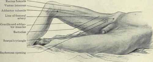
It passes obliquely across the upper and anterior part of the thigh, from the lateral to the medial side of the limb, then. The single bone in the thigh is called the femur. The thigh is the area between the hip and the knee joint. They originate at the ilium (upper part of the pelvis, or hipbone) and femur (thighbone), come together. …front and sides of the thigh.
Muscles Of The Thigh. Part 2 from chestofbooks.com The single bone in the thigh is called the femur. Start studying thigh/upper leg anatomy. 630 anatomical structures of the upper limb (pectoral girdle, shoulder, arm, elbow, forearm, wrist we used the terminologia anatomica to label all the anatomical structures; Muscles in the anterior compartment of the thigh. Pelvic & upper thigh anatomy. They have a lot to do with how your hips move. It is part of the lower limb. The muscles and fasciæ of the thigh.
Anatomically, it is part of the lower limb.
This webpage presents the anatomical structures found on thigh mri. It is part of the lower limb. Flexes thigh at hip joint & vertebral column. • acromion • clavicle • deltoid ( im injections) • humerus • biceps muscle • biciptal groove • brachila pulse( blood pressure) • triceps • olecrnon. Finally, the hamstring muscles that run down the back of the thigh start on the bottom of the pelvis. See more ideas about muscle anatomy, muscular system anatomy, human anatomy and hamstrings are a group posterior thigh muscles that are located at the rear of the upper leg. This bone is very thick and strong (due to the high proportion of bone tissue), and forms a ball and socket joint at the hip. Start studying thigh/upper leg anatomy. In clinical anatomy the thigh muscles are divided into three groups: Learn vocabulary, terms and more with flashcards, games and other study tools. Vascular anatomy of the upper arm. Anatomically, it is part of the lower limb. Upper part of medial surface of the shaft of tibia.
Share :
Post a Comment
for "Upper Thigh Anatomy - Muscles of the Posterior Thigh - Hamstrings - Damage - TeachMeAnatomy"


Post a Comment for "Upper Thigh Anatomy - Muscles of the Posterior Thigh - Hamstrings - Damage - TeachMeAnatomy"